

Carolina Panthers great Luke Kuechly promotes Q-Collar he believes extended his career
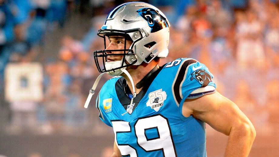
Former Carolina Panthers linebacker Luke Kuechly (59) in 2019, wearing the black Q-Collar around his neck, which he believes extended his playing career. Dannie Walls/Icon Sportswire
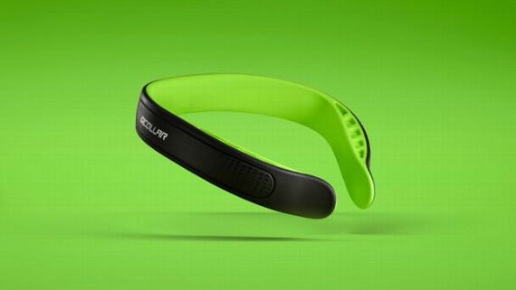
The Q-Collar was designed to reduce the chance for concussion, particularly damage from repetitive hits, and has been approved by the Food and Drug Administration (FDA). Q-Collar
INDIANAPOLIS – Luke Kuechly spent the 2021 football season hunting, fishing and traveling to beautiful wilderness spots he’d dreamed about, experiencing things he never had time for during his eight years as a Pro Bowl linebacker for the Carolina Panthers and one year as a scout for the team.
He got the same glow when he talked about shooting an elk with his dad as when he talked about the pick-six that finished off a win over the Arizona Cardinals in the 2015 NFC Championship Game.
At 30, Kuechly looks as though he could still be one of the NFL’s elite defenders, although he insists the 20 pounds he shed since retiring in January 2020 would make that tough.
“I’d get beat up,’’ Kuechly said with a laugh.
But Kuechly misses football, and after a six-month hiatus connecting with nature and himself, he’s looking for ways to again be associated with the game.
That’s why he resurfaced last week at the NFL combine, two years after his retirement. Kuechly was there to promote the Q-Collar he wore in his final three seasons after suffering his third concussion in three years in 2016 during a prime-time telecast against the New Orleans Saints.
The thin collar wraps around the neck and applies pressure on the jugular vein to increase blood volume in the skull. That creates an airbag effect around the brain that inventor Dr. David Smith and some other scientists believe reduces the chance for concussion, particularly damage from repetitive hits.
“I love the game of football,’’ Kuechly said. “If I had the opportunity to play it forever, I would have.’’
While Kuechly won’t flat-out say concussions ended his career at 28, there is no denying they were a factor.
There’s also no denying the Q-Collar he wore on an experimental basis will get more attention now that it has been approved by the Food and Drug Administration.
Kuechly believes the approval will convince more NFL players to wear the Q-Collar. The only ones who have worn it in games are Kuechly, Buffalo Bills linebacker A.J. Klein and former Pro Bowl tight end Vernon Davis.
There wasn’t much Kuechly or others could say about the collar in 2017 to convince other players to join him because it still was in the experimental stage. “Hush, hush,” as Kuechly likes to say when he’s not allowed to talk about something.
But now the future Hall of Famer wants to tell everyone about the device he believes helped extend his career.
“It’s one of those things, I trust that it works,’’ Kuechly said. “There’s a lot of other examples of it working. Did it help extend? I would hope so. Did it hurt? Absolutely not.’’
‘Luuuu-ke’ stamp of approval
Smith said he felt like a father watching his baby walk for the first time when Kuechly became the first NFL player to wear the Q-Collar in 2017. He’d seen positive results from Kuechly’s high school team, St. Xavier in Cincinnati, which had used the collar for four years.
He’d seen positive results from athletes on other high school and college teams.
But to see the collar in the NFL was a monumental step.
“The credibility Luke brings to bear, just his visibility and just the likable nature he is,’’ Smith said. “He’s just a football player’s player. That part has been a wonderful boon to having people immediately understand this just isn’t a toy.
“This is a real medical device; it’s really truly being used by the best of the best, and they’re getting huge benefits from it.’’
The NFL hasn’t officially approved the collar. A league spokesperson said only that it will be reviewed as “any technology that could have a positive effect on player safety.’’
Suzanne Williams, the vice president of sports marketing for Q30 Innovations, which produces the Q-Collar, said it’s acceptable in the NFL because it’s being promoted as a medical device, not as equipment.
The FDA approval in February 2021 said much of what Kuechly couldn’t until now.
“It’s critical,’’ Smith said. “We had a lot of back and forth with some of the best scientists in the world, helping the FDA understand all these microstructural changes we were seeing in the brain are drastically and dramatically diminished.
“Not just a little fraction. It’s dramatic. It took a while for the FDA to come around. The medical world has believed for decades that those microstructural changes are damage. The FDA finally came out and said they are damage.’’
Kuechly said he felt safer with the collar to the point where he seldom took it off as a player. He considered it a competitive edge that allowed him to play at a high level, comparable to film study or workouts.
He looked at the collar no differently than he did a mouthpiece or shoulder pads.
“Everybody is always looking for the edge, how can I be as healthy as possible?’’ said Kuechly, who is unsure what his role in football will be past promoting the collar. “That was a big reason for me [wearing] it.’’
‘Feather in the cap’
Kuechly began looking for that edge when he missed the final six games of the 2016 season after being carted off in tears following an awkward hit of Saints running back Tim Hightower. He heard from the St. Xavier High team trainer through his mother that results with the collar were good, so he tried it.
“Back then, he was like, ‘I don’t know if it works, but I’ve got nothing to lose,’ ’’ Q30 Innovations' Williams said.
Today, the science backs up what Smith and others were promoting -- that the collar helps protect the brain.
The FDA approval assessed the safety and effectiveness of the Q-Collar through several studies, including one with 284 high school football players 13 years or older. In the study, scans found no significant changes in deeper tissues of the brain in 77% of the players who wore the collar, compared with 27% who didn’t wear it.
Smith, whose son was the first to wear the collar in competition, was elated with the study results.
“I have a hard time thinking anybody that actually looked at the studies and seeing what it does wouldn’t feel the same way,’’ he said. “This is actual science.’’
There were skeptics in the science field when Kuechly first began wearing the collar, but that didn’t prevent the FDA from approving it. One expert in the world of concussion study who asked not to be identified in a 2017 ESPN.com article on the collar said “playing with blood vessels is really risky and could have negative consequences.’’
Eric Nauman, a professor of biomedical engineering and basic medical science at Purdue University, is part of the school’s Neurotrauma Group that has conducted studies on 550 football and soccer players. He, too, was skeptical in 2017.
“We actually did not pursue this one because we had concerns about the idea of pressing on the [jugular] vein, especially in an uncontrolled way,’’ he said.
The science that had Smith initially look at pressure on the jugular vein came from studying animals, particularly the woodpecker, earning him the nickname “the Bird Brain Doctor.’’
He spent nine months researching the woodpecker and other G-force-sustaining animals trying to figure out why, after repetitive blows to the head area, the bird could “fly away without a headache.’’
Smith determined that nature created internal devices to modulate and change the pressure and volume inside the cranial space to prevent concussive symptoms. He modeled the collar after that.
Kuechly was just looking for a device that would allow him to continue playing the game in which he accumulated 1,092 tackles from 2012 to 2019, more than any player in the NFL during that span.
To have Kuechly back now promoting the collar, Smith said, “is a feather in the cap.’’
“Nobody wants to wear more equipment, right?’’ Smith said. “Nobody wants to put another thing on. But this is protecting their most valid and most treasured part of your body, being your brain.
“And Luke knew that early on. To come back and say he wanted to give back and represent the collar, that’s just aces.’’
Not Peer-Reviewed, but recently generated abstracts…

TBI Future Care Today: Optimize Internal Jugular Vein Compression Collar Mitigates Histopathological Alterations after Closed Head Rotational Head Impact in Swine: A Pilot Studying Warfighter Brain Health following TBI
20200615 | 2020 Internal Jugular Vein Compression Collar Mitigates Histopathological Alterations after Closed Head Rotational Head Impact in Swine: A Pilot Study |
ABSTRACT. Recently, there has been increased concern about microstructural brain changes after head trauma. Clinical studies have investigated a neck collar that applies gentle bilateral jugular vein compression, designed to increase intracranial blood volume and brain stiffness during head trauma, which neuroimaging has shown to result in a reduction in brain microstructural alterations after a season of American football and soccer. Here, we utilized a swine model of mild traumatic brain injury to investigate the effects of internal jugular vein (IJV) compression on histopathological outcomes after injury. Animals were randomized to collar treatment (n = 8) or non-collar treatment (n = 6), anesthetized and suspended such that the head was supported by breakable tape. A custom-built device was used to impact the head, thus allowing the head to break the tape and rotate along the sagittal plane. Accelerometer data were collected for each group. Sham injured animals (n = 2) were exposed to anesthesia only. Following single head trauma, animals were euthanized and brains collected for histology. Whole slide immunohistochemistry was analyzed using Qupath software. There was no difference in linear or rotational acceleration between injured collar and non-collar animals (p > 0.05). Injured animals demonstrated higher levels of the phosphorylated tau epitope AT8 (p < 0.05) and the inflammatory microglial marker IBA1 (p < 0.05) across the entire brain, but the effect of injury was markedly reduced by collar treatment (p < 0.05) The current results indicate that internal jugular venous compression protects against histopathological alterations related to closed head trauma exposure.
Click here to LINK to the publication where you can purchase a .pdf
TBI Future Care Today: Optimizing Warfighter Brain Health following TBI
210801 | 2021 Hypocapnia Worsens Secondary Damage in Patients with Severe Traumatic Brain Injury
ABSTRACT. We hypothesized that hypocapnia (low CO2) is a risk factor for the occurrence of spreading depolarizations (SDs) in patients with sTBI. SDs are the mechanism of acute lesion development in cerebral gray matter and are a marker and mechanism of secondary injury…. Conclusions: Results demonstrate a strong independent effect of hypocapnia to worsen secondary injury after sTBI, as evidenced by intracranial monitoring of SDs. They further show that hypocapnia is common in neurointensive care, even without intentional hyperventilation. We suggest that, to optimize recovery, normocapnia should be more strictly maintained and hyperventilation should be used only in cases of life-threatening ICP elevation.
Click here to open and download PDF of the above study
Carbon Dioxide Inhalation Mitigates Functional Deficits Elicited by Blast-Induced Traumatic Brain Injury
210514 | 2021 FASB Carbon Dioxide Inhalation Mitigates Functional Deficits Elicited by Blast-Induced Traumatic Brain Injury.
ABSTRACT. Traumatic Brain Injury (TBI) is a significant public health burden affecting nearly three million individuals within the US alone annually. Large amounts of research dollars have been spent attempting to ameliorate the effects of TBI, however these efforts have failed to reduce injury rates or minimize the effects of primary injury incurred during activities such as contact sports or active military deployment…. We hypothesize that increased CO2 inhalation acting to minimize the CRV acts to minimize SLOSH and is therefore protective in the context of TBI.
Click here to open and download PDF of the above study
Peer-Reviewed Published Journal Articles
Please find a collection of peer-reviewed medical journal articles below supporting the concept of jugular compression for minimizing the risk of mild TBIs in various situations. The control numbers are the date of publication in the form YYMMDD, and the articles are presented in latest published to oldest…
Internal Jugular Vein Compression Collar Mitigates Histopathological Alterations after Closed Head Rotational Head Impact in Swine: A Pilot Study
Neuroscience, 15 June 2020, RebekahMannixabNicholas J.MorrissaGrace M.ConleyaWilliam P.MeehanIIIbcdeArthurNedderiJianhuaQiuadJamisonFloatfChristopher A.DiCesarefGregory D.Myeref
Abstract—Recently, there has been increased concern about microstructural brain changes after head trauma. Clinical studies have investigated a neck collar that applies gentle bilateral jugular vein compression, designed to increase intracranial blood volume and brain stiffness during head trauma, which neuroimaging has shown to result in a reduction in brain microstructural alterations after a season of American football and soccer. Here, we utilized a swine model of mild traumatic brain injury to investigate the effects of internal jugular vein (IJV) compression on histopathological outcomes after injury. Animals were randomized to collar treatment (n = 8) or non- collar treatment (n = 6), anesthetized and suspended such that the head was supported by breakable tape. A custom-built device was used to impact the head, thus allowing the head to break the tape and rotate along the sagittal plane. Accelerometer data were collected for each group. Sham injured animals (n = 2) were exposed to anesthesia only. Following single head trauma, animals were euthanized and brains collected for histology. Whole slide immunohistochemistry was analyzed using Qupath software. There was no difference in linear or rotational acceleration between injured collar and non-collar animals (p > 0.05). Injured animals demonstrated higher levels of the phosphorylated tau epitope AT8 (p < 0.05) and the inflammatory microglial marker IBA1 (p < 0.05) across the entire brain, but the effect of injury was markedly reduced by collar treatment (p < 0.05) The current results indicate that internal jugular venous compression protects against histopathological alterations related to closed head trauma exposure. ! 2020 IBRO. Published by Elsevier Ltd. All rights reserved.
NOTE that the researchers were from Harvard University and Cincinnati Children’s Medical Center, which are two of the top five research institutions in the Country (World?), and here’s the truly significant discovery from the study: The underlined sentence above describes the initial findings of overcoming Chronic Traumatic Encephalopathy (CTE), on which the sports world is spending hundreds of millions of dollars to find a way to block this disease from their athletes, and this study shows proof of something that worked in animals.
Abstract found in ScienceDirect. Click here to download the Abstract only.
Myer 2018 BJSM Altered brain microstructure in association with repetitive subconcussive head impacts and the potential protective effect of jugular vein compression (180903)
180903 | Myer 2018 BJSM Altered brain microstructure in association with repetitive subconcussive head impacts and the potential protective effect of jugular vein compression: a longitudinal study of female soccer athletes
ABSTRACT. Purpose To (1) quantify white matter (WM) alterations in female high school athletes during a soccer season and characterise the potential for normalisation during the off-season rest period, (2) determine the association between WM alterations and exposure to repetitive subconcussive head impacts, and (3) evaluate the efficacy of a jugular vein compression collar to prevent WM alterations associated with head impact exposure.
Click here to open and download PDF of the above study
Red Blood Cell Response to Blast Levels of Force Impartations Into Freely Moveable Fluid Surfaces Inside a Closed Container (180621)
180621 | Frontiers in Physics | David Smith, Robert Franco, Christopher A. DiCesare, Daniel K. Schneider, ChuckMcGill,QuintonD.Smith, and GregoryD.Myer
Background: Blast waves have plagued mankind for centuries, yet their interaction with blood has been largely overlooked. Recent studies of ways to mitigate traumatic brain injury (TBI) in sport have utilized slosh-reducing techniques with varying success. However, hydrodynamic principles have not been used to assess the interaction of intense blast waves and the red blood cells themselves. | Conclusions: Damage to human blood resulting from the impartation of a force similar to an IED blast was mitigated by fully containing blood within a volume and thereby reducing fluid slosh.
Click here to open and download PDF of the above study
White Matter Alterations Over the Course of Two Consecutive High-School Football Seasons and the Effect of a Jugular Compression Collar: A Preliminary Longitudinal Diffusion Tensor Imaging Study (171016)
171016 | Human Brain Mapping 00:00–00 (2017) | Weihong Yuan , Kim D. Barber Foss, Staci Thomas, Christopher A. DiCesare, Jonathan A. Dudley, Katie Kitchen, Brooke Gadd, James L. Leach, David Smith, Mekibib Altaye, Paul Gubanich, Ryan T. Galloway, Paul McCrory, Julian E. Bailes, Rebekah Mannix, William P. Meehan III, and Gregory D. Myer
Abstract: The cumulative effects of repetitive subclinical head impacts during sports may result in chronic white matter (WM) changes and possibly, neurodegenerative sequelae. In this pilot study, we investigated the longitudinal WM changes over the course of two consecutive high-school football sea- sons and explored the long-term effects of a jugular vein compression collar on these WM alterations. Diffusion tensor imaging data were prospectively collected both pre- and postseason in the two consecutive seasons. Participants were assigned into either collar or noncollar groups. Tract-based spatial statistics (TBSS) approach and region of interest-based approach were used to quantify changes in WM diffusion properties. Despite comparable exposure to repetitive head impacts, significant reductions in mean, axial, and/or radial diffusivity were identified in Season 1 in multiple WM regions in the noncollar group but not in the collar group. After an 8- to 9-month long off-season, these changes observed in the noncollar group partially and significantly reversed but also remained significantly different from the baseline. In Season 2, trend level WM alterations in the noncollar group were found but located in spatially different regions than Season 1. Last, the WM integrity in the collar group remained unchanged throughout the four time points. In conclusion, we quantitatively assessed the WM structural changes and partial reversal over the course of two consecutive high-school football seasons. In addition, the mitigated WM alterations in athletes in the collar group might indicate potential effect of the collar in ameliorating the changes against repetitive head impacts. Hum Brain Mapp 00:000–000, 2017. VC 2017 Wiley Periodicals, Inc.
Click here to open and download PDF of the above study
Internal jugular vein blood flow in the upright position during external compression and increased central venous pressure: an ultrasound study in healthy volunteers
(170515)
(170515)
170515 | Canadian Journal of Anesthesiologists | Tze Yeng Yeoh, MBChB, MMed (Anaes) . Lashmi Venkatraghavan, MD, FRCA, FRCPC . Joseph A. Fisher, MD . Massimiliano Meineri, MD
Abstract | Background External compression of the jugular veins is an effective method to increase intracranial blood volume and brain stiffness in rats and healthy volunteers. It has been reported that, on assuming an upright posture, cerebral venous drainage is distributed away from the internal jugular veins (IJVs) to the cervical venous plexus, causing complete collapse of the IJV. If so, it is not clear why external IJV compression would increase intracranial blood volume, but the latter is frequently observed in neurosurgery in the sitting position. The aim of this study was to observe the effect of external IJV compression and the Valsalva maneuver on the change in IJV cross- sectional area and IJV flow in volunteers in the upright posture. | Conclusions Compression of the internal jugular veins or an increase in intrathoracic pressure does not reduce venous drainage but actually may increase intracranial venous volume.
Click here to open and download PDF of the above study
Effect of Internal Jugular Vein Compression on Intracranial Hemorrhage in a Porcine Controlled Cortical Impact Model (170415)
170415 | JOURNAL OF NEUROTRAUMA | Brian Sindelar, Julian Bailes, Sydney Sherman, John Finan, James Stone, John Lee, Saman Ahmadian, Ying Zhou, Vimal Patel, and David Smith
Abstract: Internal jugular vein (IJV) compression has been shown to reduce axonal injury in pre-clinical traumatic brain injury (TBI) models and clinical concussion studies. However, this novel approach to prophylactically mitigating TBI through venous congestion raises concerns of increasing the propensity for hemorrhage and hemorrhagic propagation. This study aims to test the safety of IJV compression in a large animal controlled cortical impact (CCI) injury model and the resultant effects on hemorrhage. Twelve swine were randomized to placement of a bilateral IJV compression collar (CCI+collar) or control/no collar (CCI) prior to CCI injury. A histological grading of the extent of hemorrhage, both subarachnoid (SAH) and intraparenchymal (IPH), was conducted in a blinded manner by two neuropathologists. Other various measures of TBI histology were also analyzed including: b-amyloid precursor protein (b-APP) expression, presence of degenerating neurons, extent of cerebral edema, and inflammatory infiltrates. Euthanized 5h after injury, the CCI+collar animals exhibited a significant reduction in total SAH ( p = 0.024–0.026) and IPH scores ( p = 0.03-0.05) compared with the CCI animals. There was no statistically significant difference in scoring for the other markers of TBI (b-APP, neuronal degeneration, cerebral edema, or inflammatory infiltration). In conclusion, IJV compression was shown to reduce hemorrhage (SAH and IPH) in the porcine CCI model when applied prior to injury. These results suggest the role of IJV compression for mitigation of not only axonal, but also hemorrhagic injury following TBI.
Click here to open and download PDF of the above study
Protection against Blast-Induced Traumatic Brain Injury by Increase in Brain Volume (170410)
170410 | Protection against Blast-Induced Traumatic Brain Injury by Increase in Brain Volume | BioMed Research International | Ming Gu, Usmah Kawoos, Richard McCarron, and Mikulas Chavk
Blast-induced traumatic brain injury (bTBI) is a leading cause of injuries in recent military conflicts and it is responsible for an increased number of civilian casualties by terrorist attacks. bTBI includes a variety of neuropathological changes depending on the intensity of blast overpressure (BOP) such as brain edema, neuronal degeneration, diffuse axonal damage, and vascular dysfunction with neurological manifestations of psychological and cognitive abnormalities. Internal jugular vein (IJV) compression is known to reduce intracranial compliance by causing an increase in brain volume and was shown to reduce brain damage during closed impact-induced TBI. We investigated whether IJV compression can attenuate signs of TBI in rats after exposure to BOP. Animals were exposed to three 110 ± 5 kPa BOPs separated by 30 min intervals. Exposure to BOP resulted in a significant decrease of neuronal nuclei (NeuN) together with upregulation of aquaporin-4 (AQP-4), 3-nitrotyrosine (3-NT), and endothelin 1 receptor A (ETRA) expression in frontal cortex and hippocampus one day following exposures. IJV compression attenuated this BOP-induced increase in 3-NT in cortex and ameliorated the upregulation of AQP-4 in hippocampus. These results suggest that elevated intracranial pressure and intracerebral volume have neuroprotective potential in blast-induced TBI.
Click here to open and download PDF of the above study
Internal Jugular Vein Compression: A Novel Approach to Mitigate Blast Induced Hearing Injury (170400)
170400 | Otology & Neurotology: April 2017 - Volume 38 - Issue 4 - p 591–598 | Sindelar, Brian; Shinners, Michael; Sherman, Sydney; Novak, Kevin; Erickson, Kristine; Patel, Vimal; Kubilis, Paul; Smith, David; Finan, John; Bailes, Julian E.
Hypothesis: Internal jugular vein (IJV) compression before blast injury will lead to reduced risk of traumatic hearing injury following exposure to a blast injury.
Background: IJV compression and its effects on not only intracranial, but also intracochlear pressure may potentiate blast induced hearing injury, therefore, precluding its use as a prophylactic therapy for blast induced traumatic brain injury. Conclusion: This study supports the use of IJV compression in a pre-clinical model as a new prophylactic mechanism to combat blast induced hearing injury.
Click here for LINK to the above study
The Effects of External Jugular Compression Applied during High Intensity Power, Strength and Postural Control Tasks (170131)
170131 | Current Research Concussion 2017; 04(01): e23-e31 DOI: 10.1055/s-0037-1606580 | Christopher A. DiCesare, MS Kim D. Barber Foss, MS, ATC Staci Thomas, MS
Daniel K. Schneider, BS Nicholas M. Edwards, MD, MPH Gregory D. Myer, PhD
Abstract. Current strategies focused on mitigating concussion in sport have demonstrated limited effectiveness. There is a paucity of research on the optimization of intracranial brain dynamics to mitigate concussion; in the present study, we investigate a novel device that provides mild jugular vein compression and may provide adjunctive protection to protect the brain internally from concussive and sub-concussive impacts. The purpose of this study was to assess the tolerance and acceptance of this device in a population of normal, healthy adults undergoing exertion similar to that is experienced while participating in sports-related competition, while monitoring changes in their biomechanical, strength, power, and postural stability capabilities.
Click here to open and download PDF of the above study
Neck Collar with Mild Jugular Vein Compression Ameliorates Brain Activation Changes during a Working Memory Task after a Season of High School Football (170000)
170000 | Journal of Neurotrauma | Yuan W; Leach J; Maloney T; Altaye M; Smith D; Gubanich PJ; Barber Foss KD; Thomas S; DiCesare CA; Kiefer AW; Myer GD
Emerging evidence indicates that repetitive head impacts, even at a sub-concussive level, may result in exacerbated or prolonged neurological deficits in athletes. This study aimed to: 1) quantify the effect of repetitive head impacts on the alteration of neuronal activity based on functional magnetic resonance imaging (fMRI) of working memory after a high school football season; and 2) determine whether a neck collar that applies mild jugular vein compression designed to reduce brain energy absorption in head impact through "slosh" mitigation can ameliorate the altered fMRI activation during a working memory task. Participants were recruited from local high school football teams with 27 and 25 athletes assigned to the non-collar and collar group, respectively. A standard N-Back task was used to engage working memory in the fMRI at both pre- and post-season. The two study groups experienced similar head impact frequency and magnitude during the season (all p > 0.05). fMRI blood oxygen level dependent (BOLD) signal response (a reflection of the neuronal activity level) during the working memory task increased significantly from pre- to post-season in the non-collar group (corrected p < 0.05), but not in the collar group. Areas displaying less activation change in the collar group (corrected p < 0.05) included the precuneus, inferior parietal cortex, and dorsal lateral prefrontal cortex. Additionally, BOLD response in the non-collar group increased significantly in direct association with the total number of impacts and total g-force (p < 0.05). Our data provide initial neuroimaging evidence for the effect of repetitive head impacts on the working memory related brain activity, as well as a potential protective effect that resulted from the use of the purported brain slosh reducing neck collar in contact sports.
Click here to open and download PDF of the above study
Current state of concussion prevention strategies: a systematic review and meta-analysis of prospective, controlled studies (161223)
161223 | British Journal of Sports Medicine | Schneider DK, et al. Br J Sports Med 2016;0:1–11. doi:10.1136/bjsports-2015-095645 | Daniel K Schneider, Ravi K Grandhi, Purnima Bansal, George E Kuntz IV, Kate E Webster, Kelsey Logan, Kim D Barber Foss, Gregory D Myer
ABSTRACT | Objective | The aim of the current review was to systematically identify, evaluate and synthesise trials that examine concussion prevention via equipment, educational programmes and training programmes. | Conclusions | Prospective controlled studies indicate that certain protective equipment may prevent superficial head injury, but these items are suboptimal for concussion prevention in sport.
Click here to open and download PDF of the above study
The effects of external Jugular compression applied during head impact exposure on longitudinal changes in Brain neuroanatomical and neurophysiological Biomarkers: a Preliminary investigation (160606)
160606 | Frontiers in Neurology | June 2016 | Volume 7 | Article 74 | Gregory D. Myer, Weihong Yuan, Kim D. Barber Foss, David Smith, Mekibib Altaye, Amit Reches, James Leach, Adam W. Kiefer, Jane C. Khoury, Michal Weiss, Staci Thomas, Chris Dicesare, Janet Adams, Paul J. Gubanich, Amir Geva, Joseph F. Clark, William P. Meehan III, Jason P. Mihalik and
Darcy Krueger
Objectives: Utilize a prospective in vivo clinical trial to evaluate the potential for mild neck compression applied during head impact exposure to reduce anatomical and physiological biomarkers of brain injury. Conclusion: Group differences in the longitudinal changes in both neuroanatomical and electrophysiological measures, as well as the correlation between the measures, provide initial evidence indicating that mild jugular vein compression may have reduced alterations in the WM response to head impacts during a competitive hockey season. The data indicate sport-related alterations in WM microstructure were ameliorated by application of jugular compression during head impact exposure. These results may lead to a novel line of research inquiry to evaluate the effects of protecting the brain from sports-related head impacts via optimized intracranial fluid dynamics.
Click here to open and download PDF of the above study
Analysis of head impact exposure and brain microstructure response in a season-long application of a jugular vein compression collar: a prospective, neuroimaging investigation in American football (160521)
160521 | British Journal of Sports Medicine | Myer GD, et al. Br J Sports Med 2016;0:1–11. doi:10.1136/bjsports-2016-096134 | Gregory D Myer, Weihong Yuan, Kim D Barber Foss, Staci Thomas, David Smith, James Leach, Adam W Kiefer, Chris Dicesare, Janet Adams, Paul J Gubanich, Katie Kitchen, Daniel K Schneider, Daniel Braswell, Darcy Krueger, Mekibib Altaye
ABSTRACT | Background | Historical approaches to protect the brain from outside the skull (eg, helmets and mouthpieces) have been ineffective in reducing internal injury to the brain that arises from energy absorption during sports- related collisions. We aimed to evaluate the effects of a neck collar, which applies gentle bilateral jugular vein compression, resulting in cerebral venous engorgement to reduce head impact energy absorption during collision. Specifically, we investigated the effect of collar wearing during head impact exposure on brain microstructure integrity following a competitive high school American football season. Conclusions | The data evaluated in the current project indicate that the device is safe during high intensity and dynamic postural stabilization exercise and does not alter normal physical or neuromuscular capabilities during physical activity.
Click here to open and download PDF of the above study
MR Elastography Can Be Used to Measure Brain Stiffness Changes as a Result of Altered Cranial Venous Drainage During Jugular Compression (151000)
151000 | American Journal of Neurological Research | American Journal of Neuroradiology October 2015, 36 (10) 1971-1977; DOI: https://doi.org/10.3174/ajnr.A4361 | A. Hatt, S. Cheng, X K. Tan, R. Sinkus, and L.E. Bilston
ABSTRACT | BACKGROUND AND PURPOSE: Compressing the internal jugular veins can reverse ventriculo-megalyinthe syndrome of inappropriately low pressure acute hydrocephalus, and it has been suggested that this works by “stiffening” the brain tissue. Jugular compression may also alter blood and CSF flow in other conditions. We aimed to understand the effect of jugular compression on brain tissue stiffness and CSF flow. CONCLUSIONS: Jugular compression influences cerebral CSF hydrodynamics in healthy subjects and can increase brain tissue stiffness, but the magnitude of the stiffening depends on the percentage of cranial blood draining through the internal jugular veins during compression—that is, subjects who maintain venous drainage through the internal jugular veins during jugular compression have stiffer brains than those who divert venous blood through alternative pathways. These methods may be useful for studying this phenomenon in patients with the syndrome of inappropriately low-pressure acute hydrocephalus and other conditions.
Click here to open and download PDF of the above study
The effect of jugular vein compression on cerebral hemodynamics in healthy subjects (150700)
150700 |Joseph A. Fisher M.D., James Duffin PhD., David Mikulis M.D., Olivia Sobczyk MSc., PhD (candidate)
Introduction. As upright animals, the human brain is high above the level of the heart resulting in low intracranial venous pressures. Indeed, air can be sucked into the venous sinuses during neurosurgery. This low pressure also indicates that there is room for the vessels to expand, or the presence of a capacitance for volume. It has been recently proposed that intracranial volume being less than skull volume allows the brain to be to be mobile inside the skull. In the presence of head trauma the brain may move relative to, and collide with, the skull (“rattle”) or be internally deformed by pressure waves (“slosh”), resulting in traumatic brain injury. Both mechanisms can be mitigated, or eliminated by increasing the intra-cranial compartment volume, forcing all parts of the brain and skull to move as a unit. In addition, if the intracranial volume fills the skull, the brain will conduct blast energy waves through with minimal energy absorption by the brain, avoiding tissue displacement and shear. It has been proposed that a way to expand the intracranial volume is to fill it with venous blood. Since the brain blood flow is large, a small degree of resistance to drainage will quickly fill the cerebrovascular compliance. The major route of venous drainage in humans is via the internal jugular veins (IJV). In contrast, most quadrupeds have their head at near heart level and the vertebral venous plexi are the main venous outflow conduits (Lavoie et al., 2008). Therefore, studies of the effects of jugular venous compression on brain blood flow and intracranial blood distribution must be performed in humans. Summary of findings: Compression of the internal jugular veins in supine healthy volunteers… | 1) ...increases in blood volume in the skull with venous blood distributed particularly to the large venous sinuses. A small increase in BOLD signal seen in the middle cerebral artery and cerebellum indicate the possibility of a slightly increased cerebral blood flow or the accumulation of arterial blood diffusely as well. | 2) …does not change the level or distribution of the resting blood flow in the brain. | 3) ...does not affect the response of the brain blood flow to hypercapnia. This implies that it would not affect the brain blood flow in response to an increase in metabolic demand. | 4) …does not reduce the CBF if applied during an increase in demand, as occurs in this study during hypercapnia. This finding provides confidence that the brain would be able to maintain CBF if the collar is applied under other high flow demand states such as exercise.
Click here to open and download PDF of the above study
Altitude Modulates Concussion Incidence: Implications for Optimizing Brain Compliance to Prevent Brain Injury in Athletes (140509)
140509 | The Orthopaedic Journal of Sports Medicine | David W. Smith, MD, Gregory D. Myer, Dustin W. Currie, MPH, R. Dawn Comstock, PhD, Joseph F. Clark, PhD, and Julian E. Bailes, MD
Background: Recent research indicates that the volume and/or pressure of intracranial fluid, a physiology affected by one’s altitude (ie, elevation above sea level), may be associated with the likelihood and/or severity of a concussion. The objective was to employ an epidemiological field investigation to evaluate the relationship between altitude and concussion rate in high school sports. | Conclusion: The results of this epidemiological investigation indicate increased physiological responses to altitude may be associated with a reduction in sports-related concussion rates, especially in collision sports. Future research that focuses on the potential prophy- lactic effect of optimizing outflow impedance and thus reduction of intracranial compliance (a ‘‘tighter fit’’) in humans is warranted to determine the most effective approaches to mitigate sport-related concussion, especially in football players.
Click here to open and download PDF of the above study
Rates of concussion are lower in National Football League games played at higher altitudes (140100)
140100 | Journal of Orthopaedic and Sports Physical Therapy 2014, 44 (3): 164-72 | GREGORY D. MYER, PhD, FACSM, CSCS*D • DAVID SMITH, MD • KIM D. BARBER FOSS, MS, ATC, LAT CHRISTOPHER A. DICESARE, MS, CSCS • ADAM W. KIEFER, PhD • ADAM M. KUSHNER, BS, CSCS STACI M. THOMAS, MS • HEIDI SUCHAREW, PhD • JANE C. KHOURY, PhD
OBJECTIVE: To investigate the relationship between altitude and concussion rate in the National Football League (NFL). Because of the physiologic responses that occur during acclimatization to altitude, it was hypothesized that games played on fields at a higher altitude would have reduced concussion rates compared to games played on fields at a lower altitude. CONCLUSION: The results of this epidemiological investigation indicate that increased altitude was associated with a reduction in the odds of a sport-related concussion in NFL athletes. The reported relationship of concussion incidence and field elevation should be further investigated, and, if verified, further work will be needed to understand why that relationship exists.
Click here to open and download PDF of the above study
Effect of slosh mitigation on histologic markers of traumatic brain injury (121200)
121200 | Journal of Neurosurgery / Volume 117 / December 2012 | RYAN C. TURNER, B.S., ZACHARY J. NASER, B.A., JULIAN E. BAILES, M.D., DAVID W. SMITH, M.D., JOSEPH A. FISHER, M.D., AND CHARLES L. ROSEN, M.D., PH.D.
Object. Helmets successfully prevent most cranial fractures and skull traumas, but traumatic brain injury (TBI) and concussions continue to occur with frightening frequency despite the widespread use of helmets on the athletic field and battlefield. Protection against such injury is needed. The object of this study was to determine if slosh mitigation reduces neural degeneration, gliosis, and neuroinflammation.
Click here to open and download PDF of the above study
Internal Jugular Vein Compression Mitigates Traumatic Axonal Injury in a Rat Model by Reducing the Intracranial Slosh Effect (110905)
110905 | Neurosurgery, Volume 70, Issue 3, 1 March 2012, Pages 740–746 | David W. Smith, MD Julian E. Bailes, MD Joseph A. Fisher, MD Javier Robles, MD Ryan C. Turner, BS James D. Mills, MD
Abstract | Background: Traumatic brain injury (TBI) remains a devastating condition for which extracranial protection traditionally has been in the form of helmets, which largely fail to protect against intracranial injury. Objective: To determine whether the pathological outcome after traumatic brain injury can be improved via slosh mitigation by internal jugular vein (IJV) compression. Conclusion: Using a standard acceleration-deceleration laboratory model of mild traumatic brain injury, we have shown successful prevention of axonal injury after IJV compression as indicated by immunohistochemical staining of amyloid precursor protein. We argue that IJV compression reduces slosh-mediated brain injury by increasing intracranial blood volume, which can be indirectly measured by intracranial and intraocular pressures.
Click here to open and download PDF of the above study
